Introduction to Levels of Organisation
Living things are incredibly organised! From the tiniest parts inside cells to whole organisms, there's a clear hierarchy. Think of it like Russian dolls - each level fits inside the next, getting bigger and more complex. This organisation helps living things work efficiently and stay alive.
Key Definitions:
- Organelle: A tiny structure inside a cell that has a specific job, like the nucleus or mitochondria.
- Cell: The basic unit of life - the smallest thing that can be called 'living'.
- Tissue: A group of similar cells working together to do the same job.
- Organ: Different tissues working together to perform a specific function.
- Organ System: Several organs working together, like your digestive system.
- Organism: A complete living thing made up of all the organ systems.
🔍 The Hierarchy in Action
Imagine eating an apple. Your teeth (organs) break it down, your stomach (organ) digests it and muscle cells (cells) in your intestines move it along. The mitochondria (organelles) in those muscle cells provide the energy needed. Every level works together!
Organelles - The Cell's Workforce
Organelles are like tiny factories inside cells, each with their own special job. Just like how a school has different rooms for different subjects, cells have different organelles for different functions. Let's meet the key players!
Essential Organelles and Their Functions
Every organelle has evolved to do a specific job perfectly. Understanding what each one does helps us understand how cells work as a whole.
🏠 Nucleus
The cell's control centre. Contains DNA (genetic instructions) and controls all cell activities. Think of it as the headteacher of the cell!
⚡ Mitochondria
The powerhouses! They break down glucose to release energy (ATP) for the cell. More active cells have more mitochondria.
🔧 Ribosomes
Protein factories. They read instructions from DNA and build proteins. Found floating freely or attached to rough ER.
📦 Endoplasmic Reticulum
Transport network. Rough ER (with ribosomes) makes proteins. Smooth ER (no ribosomes) makes lipids and detoxifies.
🚚 Golgi Apparatus
The post office! Modifies, packages and ships proteins from the ER to where they're needed in the cell.
🗑 Lysosomes
Recycling centres. Contain digestive enzymes that break down worn-out organelles and harmful substances.
Amazing Fact!
A single liver cell can contain over 2,000 mitochondria! This is because liver cells work incredibly hard, processing toxins and making proteins, so they need loads of energy.
Plant vs Animal Cells - Spot the Difference
While plant and animal cells share many organelles, plants have some special extras that help them make their own food and stay upright. These differences reflect their different lifestyles!
Plant-Only Organelles
Plants are amazing because they can make their own food from sunlight. They also need to stay rigid without bones. Special organelles help them do both!
Case Study: Palisade Leaf Cells
Palisade cells in leaves are packed with chloroplasts (sometimes 50+ per cell!) and arranged in neat columns. This maximises light absorption for photosynthesis. They're like solar panels optimised for capturing sunlight!
Structure Meets Function - Perfect Partnerships
In biology, structure and function go hand in hand. The shape and features of organelles are perfectly suited to their jobs. It's like how a hammer's shape makes it perfect for hitting nails!
Examples of Structure-Function Relationships
Let's explore how the design of organelles makes them incredibly efficient at their specific roles.
⚡ Mitochondria Structure
Have folded inner membranes called cristae. These folds massively increase surface area for energy production reactions. More surface area = more energy!
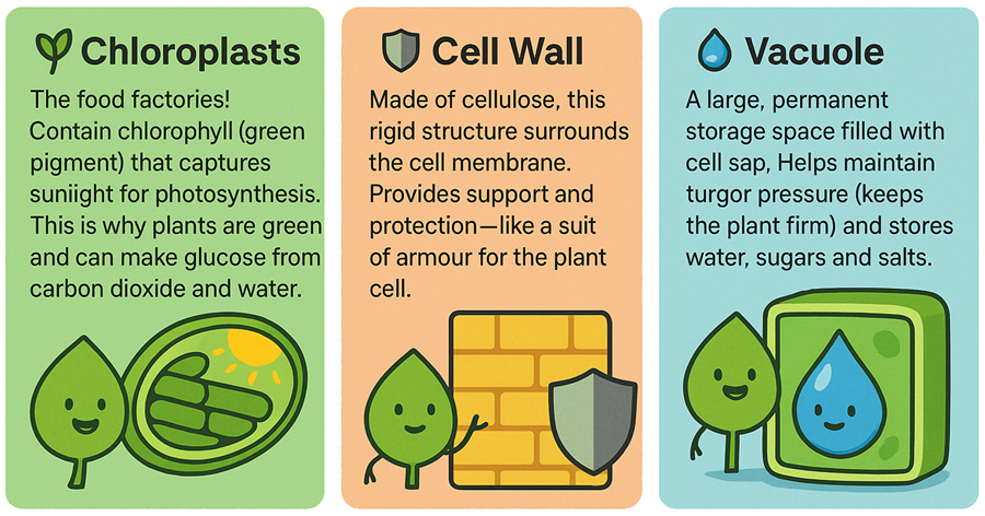
🌱 Chloroplast Structure
Contain stacks of membranes called thylakoids where light reactions happen. The large surface area captures maximum light for photosynthesis.
Specialised Cells - Experts at Their Jobs
Not all cells are the same! Different cells have different jobs, so they develop special features. It's like how different people have different careers and develop different skills.
Examples of Cell Specialisation
Specialised cells show us how structure perfectly matches function. Each type has evolved specific features for their role.
🩸 Red Blood Cells
No nucleus (more room for haemoglobin), biconcave shape (more surface area), flexible (squeeze through capillaries).
🌱 Root Hair Cells
Long projections increase surface area for water absorption. Lots of mitochondria provide energy for active transport.
⚡ Muscle Cells
Packed with mitochondria for energy, contain protein filaments that can contract, elongated shape allows shortening.
Case Study: Sperm Cells
Sperm cells are swimming champions! They have a streamlined head, a tail for swimming and mitochondria packed in the middle section for energy. Everything about their structure helps them reach and fertilise an egg cell.
Putting It All Together
Understanding levels of organisation helps us see how life works from the smallest to the largest scale. Every organelle, cell, tissue and organ has evolved to work perfectly together. It's like a massive, incredibly complex machine where every part has a purpose!
Remember: organelles work together in cells, cells work together in tissues, tissues work together in organs, organs work together in systems and systems work together in organisms. This organisation is what makes life possible and efficient.